partial coverage alignment failure - follow-up

AFNI version info (afni -ver):
Precompiled binary linux_ubuntu_16_64: May 23 2025 (Version AFNI_25.1.11 'Maximinus')

Dear AFNI,
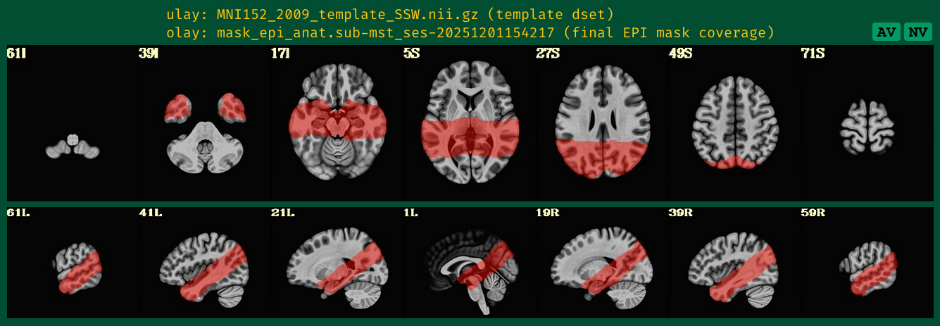
I am following up on the forum thread partial coverage alignment failure from January 2024 that seemed to stop because of technical issues. I am having a very similar issue since I am also trying to acquire a few BOLD fMRI slices including the hippocampus.I have only tried afni_proc so far, both on MNI (i.e. with tlrc option) and on subject space (ithout tlrc option). Unless you think it would be better to focus on MNI script, I will focus on T1w space since I am running only one participant to tune the parameters for now.

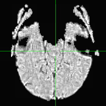
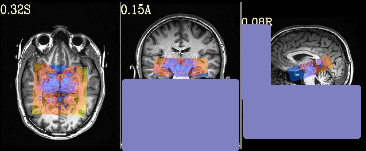
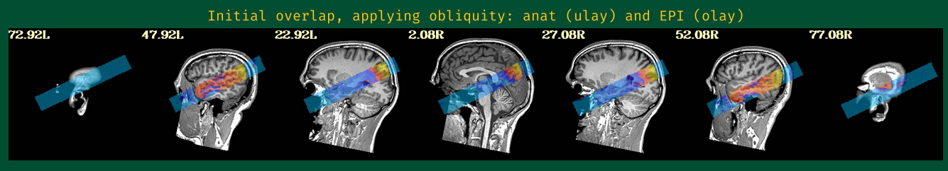
This is the beginning of output. When checking the volumes in original space, the images when applying obliquity look almost correct. That's what I acquired.

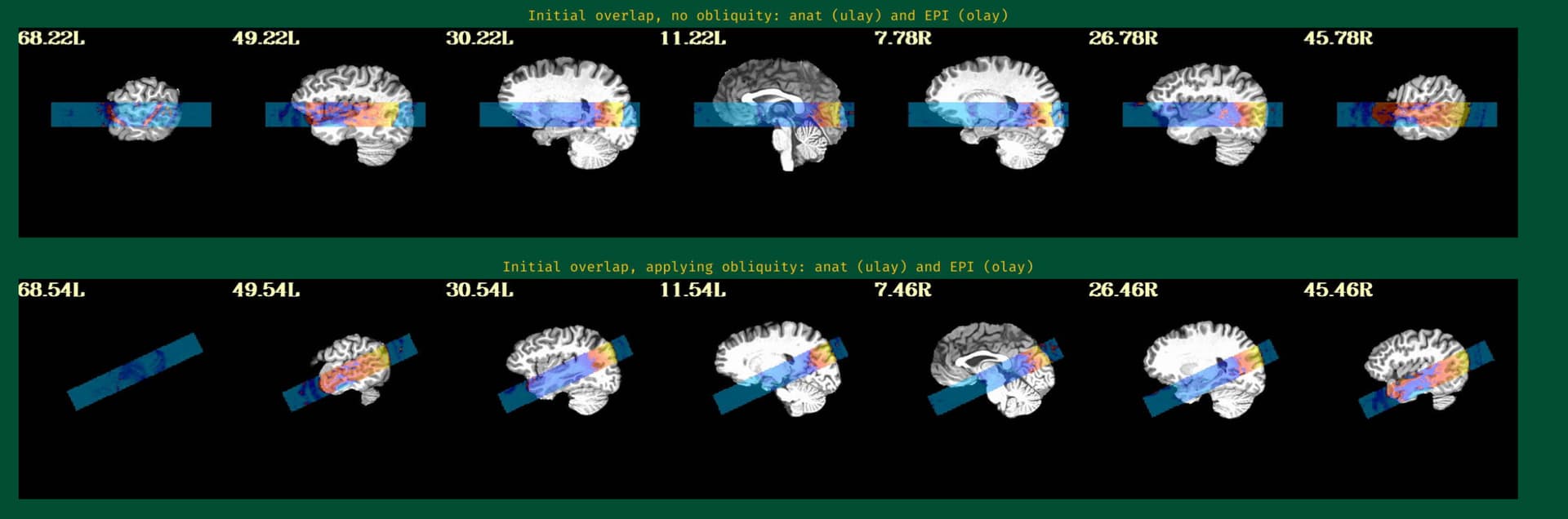
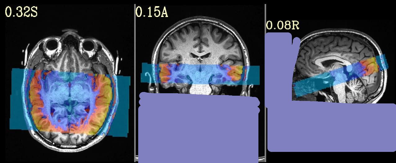
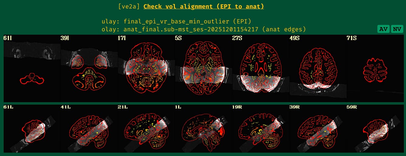
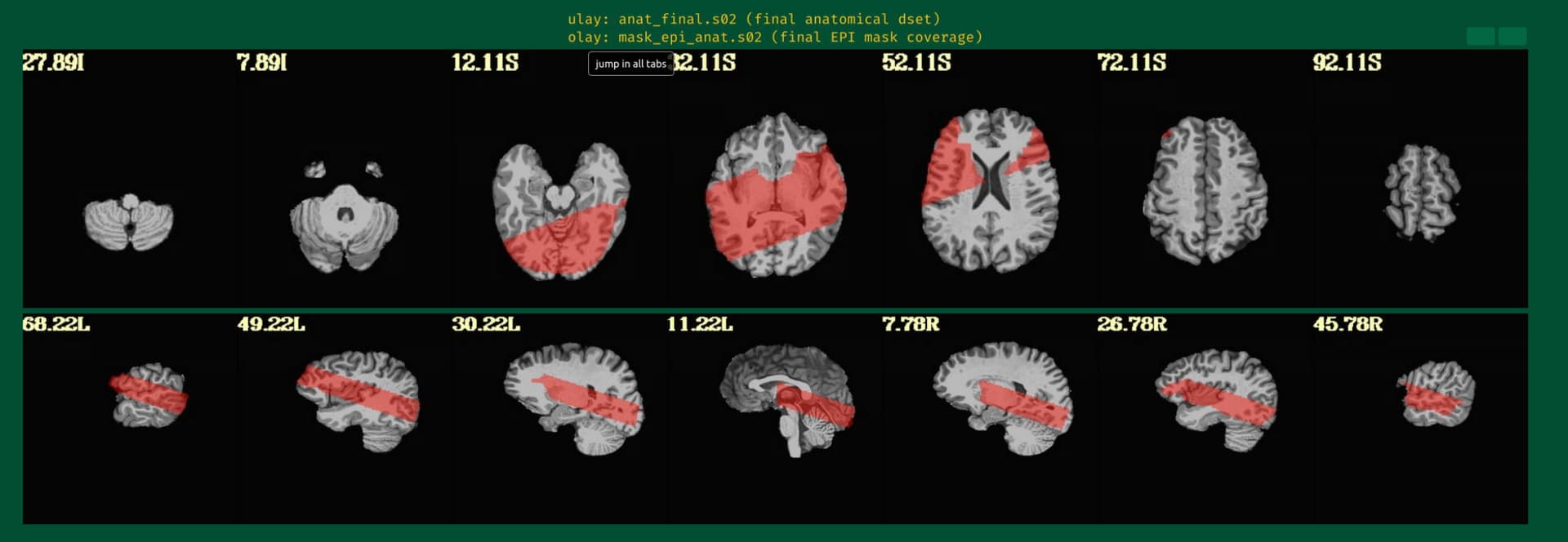
But when checking for EPI to Anat alignment, it looks like follows (almost like applying re-obliquing twice):

As an answer to the previous thread I am following, this is the output of

3dinfo -obliquity -prefix anat_final.s02+orig final_epi_vr_base_min_outlier+orig 
9.366	                    anat_final.s02
0.000	     final_epi_vr_base_min_outlier

and

@djunct_overlap_check                                           \
    -ulay    anat_final.s02+orig                                          \
    -olay    final_epi_vr_base_min_outlier+orig                                          \
    -prefix  img_olap

returned

# ulay         = anat_final.s02+orig
# ulay_is_obl  = 1
# ulay_obl_ang = 9.366
# mat44 Obliquity Transformation ::
      0.995392      0.007651     -0.095584     -11.890205
     -0.020267      0.991080     -0.131722     -12.362091
      0.093724      0.133052      0.986668     -28.940544
# 
# olay         = final_epi_vr_base_min_outlier+orig
# olay_is_obl  = 1
# olay_obl_ang = 9.366
# mat44 Obliquity Transformation ::
      1.000000      0.000000      0.000000      -0.000008
      0.000000      1.000000      0.000000       0.000000
      0.000000      0.000000      1.000000       0.000000

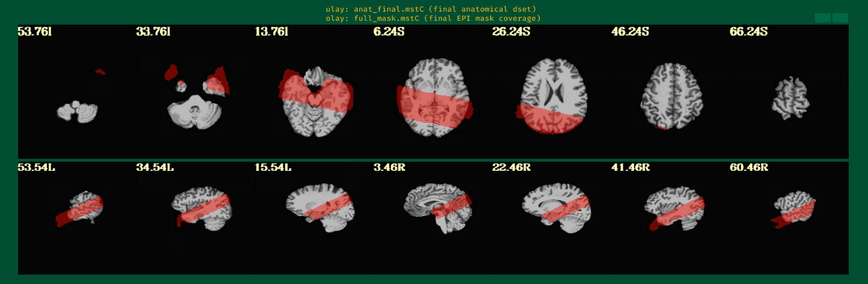
with following images:

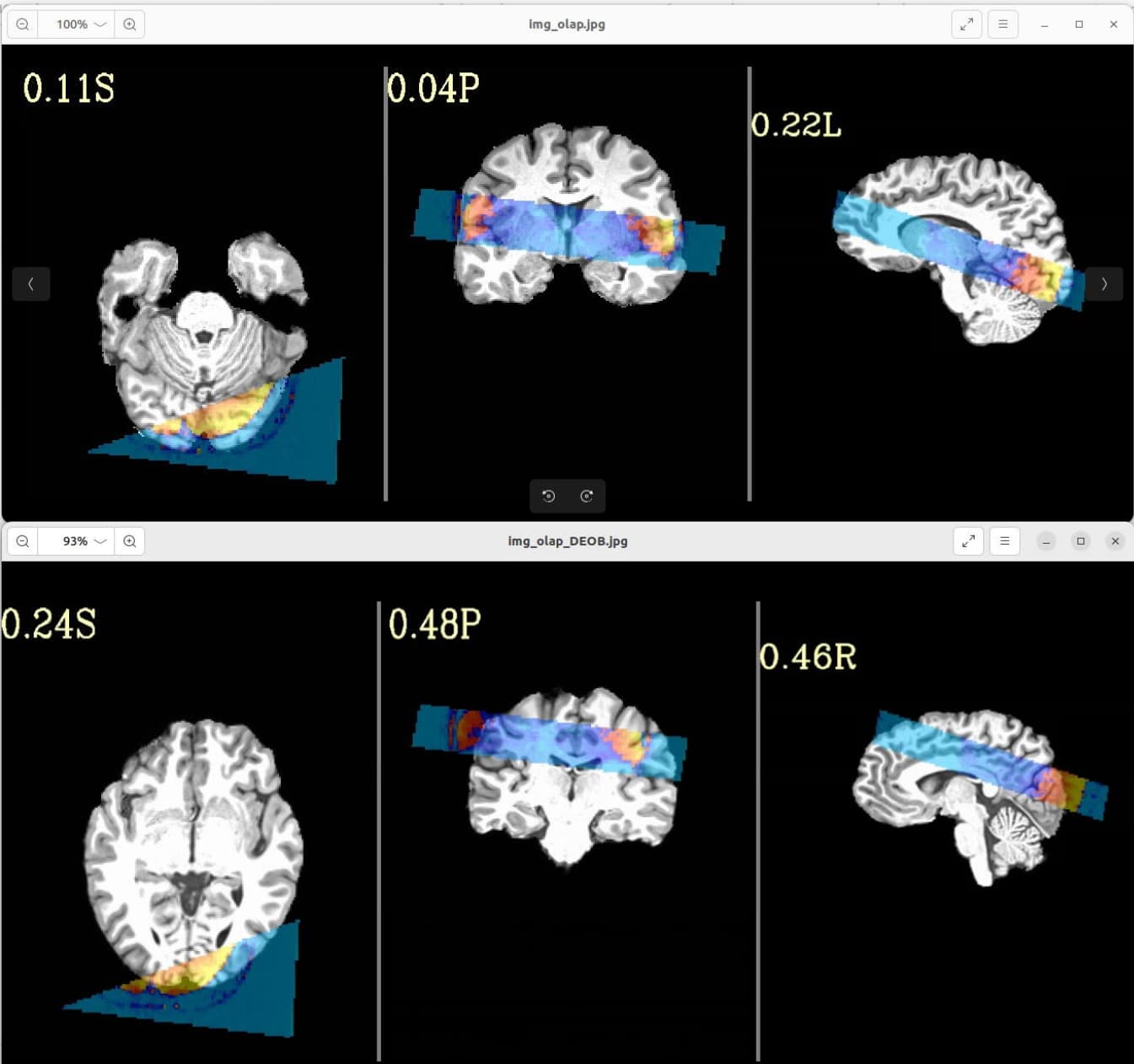
Where the DEOB doesn't seem to be correct.

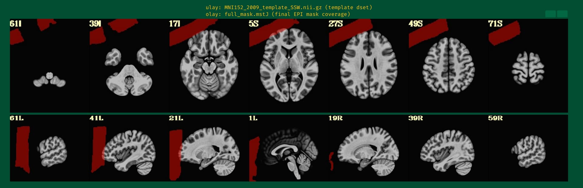
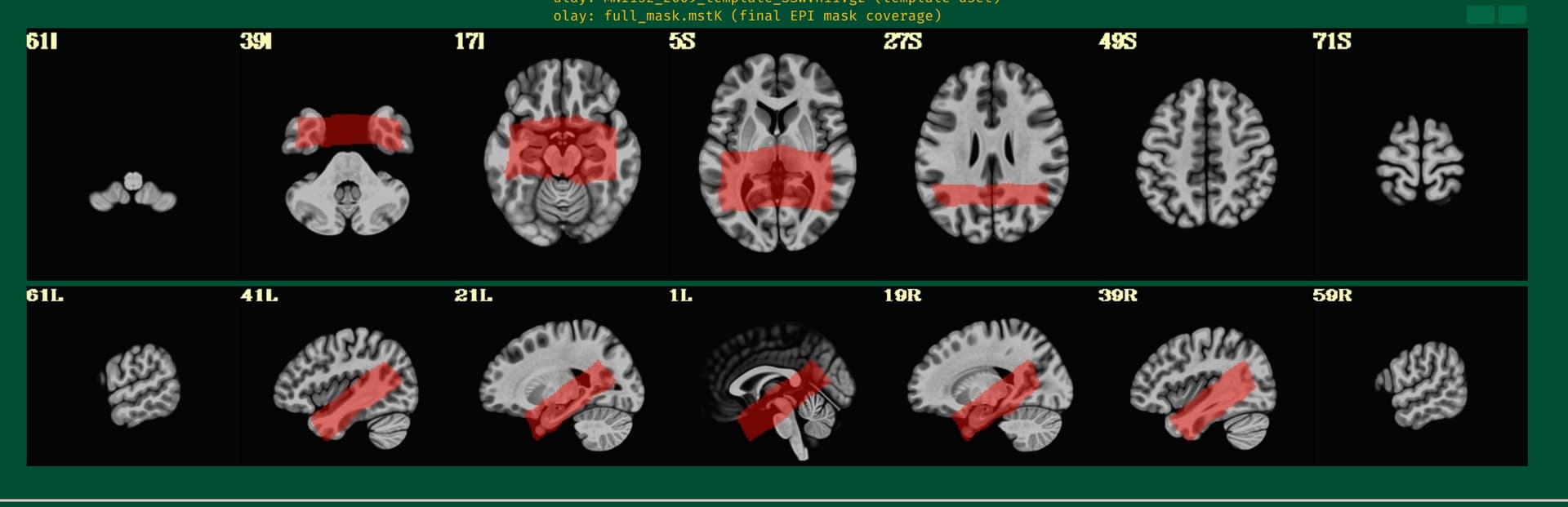
Is there a possibility to include a full brain volume (which I acquired on many more slices and totally aligned with the time series focused on hippocampus) in the afni_proc command or have I done any mistake in my command which follows?

afni_proc.py                                                         \
            -subj_id                  s02 \
            -out_dir			$subdir/S02e.results	\
            -copy_anat $subdir/o.ssw_sub-mst/anatSS.sub-mst.nii \
            -anat_has_skull           no                                     \
            -anat_follower anat_w_skull anat $subdir/sub-mst_T1w.nii.gz \
            -dsets $subdir/sub-mst_task-cmrr_run-01_bold.nii.gz \
            -blocks                   align volreg mask blur     \
                                      scale regress                          \
            -radial_correlate_blocks  tcat volreg                            \
            -tcat_remove_first_trs    0                                      \
            -align_unifize_epi        local                                  \
            -align_opts_aea           -cost lpc+ZZ                           \
                                      -giant_move                            \
                                      -check_flip                            \
            -volreg_align_to          MIN_OUTLIER                            \
            -volreg_align_e2a                                                \
            -volreg_compute_tsnr      yes                                    \
            -mask_epi_anat            yes                                    \
            -blur_size                3.0                                    \
            -regress_stim_times       $subdir/rep.1D           \
            				$subdir/new.1D         \
            -regress_stim_labels      rep new                                \
            -regress_basis_multi      'TENT(0,16,7)' 'TENT(0,16,7)'          \
            -regress_opts_3dD         -jobs 8                                \
                                      -gltsym 'SYM: rep - new'               \
                                      -glt_label 1 r-n                       \
            -regress_motion_per_run                                          \
            -regress_censor_motion    0.3                                    \
            -regress_censor_outliers  0.05                                   \
            -regress_3dD_stop                                                \
            -regress_reml_exec                                               \
            -regress_compute_fitts                                           \
            -regress_make_ideal_sum   sum_ideal.1D                           \
            -regress_est_blur_epits                                          \
            -regress_est_blur_errts                                          \
            -regress_run_clustsim     no                                     \
            -html_review_style        pythonic                               \
            -execute

Thanks!
Jonathan

hi there Jonathan-

i'm sure you'll get great feedback from the AFNI developers themselves, but since i've done so much AFNI work with small FOVs (and know how super frustrating it can be) i'll chime in. it looks to me like the align_epi_anat.py defaults are throwing your FOV out of registration. i'd like to share my experiences and advice when this happens to me.

First, get out of afni_proc for a second to speed up the troubleshooting. zero in on this alignment issue with align_epi_anat.py, the program afni_proc calls.

Second, align_epi_anat.py (aea) can be thought of a wrapper around the real workhorse 3dAllineate. i think you should refine your aea command with some deeper constraints to 3dAllineate. there are many of these: -maxrot, -maxshf, -maxscl etc.. you might also want to constrain an initial alignment as rigid body ("-warp shift_rotate").

Third, when you've found a set of parameters that work (and there are many GUI ways to check alignment carefully; you don't necessarily need the afni_proc html images for that), you can place the parameter constraints back into your afni_proc command with something like:

            -align_opts_aea -cost lpc+ZZ -check_flip \
			-Allineate_opts -warp shift_rotate -maxrot 5

this process may be a little more time consuming than testing aea's built-in options like -big_move, -giant_move, -large_move etc, but i've found it's quite flexible and robust to wrangling the most difficult datasets. hope this helps and let me know if you have further questions.

-Sam

Dear Sam,

Thank you for your answer.

I guess the align_epi_anat.py that has to be tuned is the following in the proc file generated by afni_proc:

align_epi_anat.py -anat2epi -anat anatSS.sub-mst+orig  \
       -suffix _al_junk                                \
       -epi vr_base_min_outlier_unif+orig -epi_base 0  \
       -epi_strip 3dAutomask                           \
       -anat_has_skull no                              \
       -cost lpc+ZZ -giant_move -check_flip            \
       -volreg off -tshift off

since the "al_junk" file in ouput is misaligned when I check the registration.

I am dealing with 3dAllineate right now tuning -maxrot, -maxshl, etc., but so far I am not succesful. I will still try a little more.

This being said, when I am looking at the vr_base_min_outlier_unif (picture below), I wonder if the non brain parts are the origin of the problem:

Jonathan

you may also want to try removing "-align_unifize_epi" from your original afni_proc to retain tissue contrast in the vr_base

Partial data like this can be difficult, especially with strong gradients and little structure and tissue contrast. I will add to Sam's recommendations to try a couple more things:

  1. Use -partial_xxx options to control the cmass moves.
  2. Remove -giant_move because they start out close after the deobliquing step.
  3. Tweak align_epi_anat.py options with other cost functions like nmi or lpa+ZZ for low tissue contrast data. Remove the automask option here too.
  4. You can use the full brain EPI dataset instead (if I understand correctly that you do have one) as long as it's aligned with a particular EPI subbrick volume. Select the full brain align_epi_ext_dset. Select the volreg base for motion correction specifically with the options -volreg_align_to, -volreg_base_dset and -volreg_base_ind.
1 Like

Well, I think I might have something that will help navigate dealing with obliquity and the initial spatial overlap better, which should in turn help the alignment itself. Let me see if I can finally finish it and put it into the build today (or thereabouts...).

--pt

1 Like