AFNI version info (afni -ver): AFNI_24.0.12 'Caracalla'
Hi AFNI experts!
I was analyzing some new data that came in on one of my participants and noticed the scans looked a bit odd... specifically, the anatomical images appear fuzzy and in the coronal view you can see how it looks like an image is shifted on top of the other.
Have any of you run into scans that look like this before?
The only thing I've done to this data so far is convert it from dicom to .BRIK and .HEAD files using DIMON, but I believe this issue occurred while the data was being collected in the scanner.
Thank you!
Howdy-
That is an anatomical T1w scan, and I would guess odd behavior/blurriness that is the result of subject motion during acquisition. It appears to have some ringing in the structure---a hallmark of motion during anatomical dataset acquisition. It might be a little more apparent if you show this dataset as an overlay on itself, actually, because the ringing might be clearer, as well as structures outside the brain that aren't really visible here.
--pt
Hi Paul,
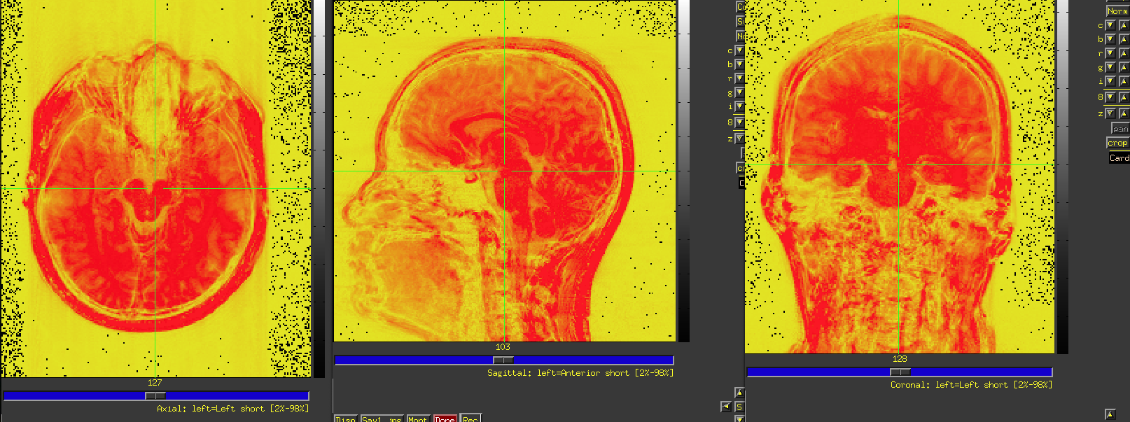
Thanks for getting back to me so quickly! I overlayed the image on top of itself, however I am not quite sure what I should be looking for to identify ringing on the scans. Here are the scans overlayed on top of one another.
I was expecting to see some structures appearing outside the head. I think I can see some faint ones along the anterior-posterior axis---see how the orange/red faintly appears in lines emanating from the brain? If you hit the "Pos" button beneath the color bar, then the min colorbar value would be 0, and more colorbar range would be used across the FOV (because anatomical dsets are usually positive-valued). That might show it a bit more clearly, but I think that is what is happening.
--pt
I think you're right and it has to do with participant motion. Thank you so much!